Infinity Optical Systems

Infinity optics refers to the concept of a beam path with parallel rays between the objective and the tube lens of a microscope. Flat optical components can be brought into this infinity space without influencing image formation, which is critical for the utilization of contrast methods such as DIC or fluorescence. Modern microscopy techniques require the addition of multiple optical instruments, such as light sources or laser devices, into the infinite light path. Different approaches to fulfill this need have emerged and are described here.Since the invention of glass by the Romans in the first century, people recognized that a magnification effect was created by round-shaped pearls of glass. Later on, this effect was scientifically investigated and further developed, resulting in the simple magnification glasses of the 16th and 17th centuries, e.g., those invented by Hans and Zacharias Jansen or Anton van Leeuwenhoek. Historically, this was microscopy’s hour of the birth. By definition, the microscope is an instrument that magnifies objects that are not normally resolvable by the human eye, and so these single lens tools were already – microscopes. Nowadays, we think of something different when we talk about a microscope. This is due to the fact that people soon realized that the combination of two separate lenses (or lens systems) in a row is a more effective visual tool than a single lens. To describe such a set-up, the term compound microscope was created. Compound microscopes consist of an objective that is magnifying the specimen and an eyepiece (respectively two), magnifying the image produced by the objective.
Introduction of infinity optics
The distance between the objective shoulder and the ocular shoulder is called mechanical tube length. For standardization, this value was set to 160 mm by the Royal Microscopical Society in the 19th century. Over the years, this design turned out to have some drawbacks. Adding additional optical elements into the light path, such as prisms for differential interference contrast (DIC), polarizers, etc., changed the effective tube length and introduced aberrations, which had to be accepted, or corrected with the addition of other hardware components. For this reason, the microscope manufacturer Reichert started to experiment with so-called infinite optics in the 1930s, and this technology was later adopted by all other microscope companies. Objectives of these infinite optical systems project a specimen image to infinity, meaning that all light rays derived from a single point of the specimen exit the objective in a parallel way. Those rays within the center of the specimen (and the objective) run parallel to the optical axis. Those outside the center of the specimen run parallel to each other, but not to the optical axis. The virtual image that an infinity-corrected objective produces has to be captured by an additional lens – the tube lens – to bring it to the front focal point of the eyepiece lens. This approach enables the addition of optical instruments such as DIC prisms into the infinity space between the objective and the tube lens without influencing image quality. Neither location nor the focal point of the image is altered. Advantages of infinity optics. Several contrast methods require the introduction of special optical components into the microscope’s light path. For example, prisms and polarizers for DIC, or dichroics and filters for fluorescence microscopy, are indispensable for the relevant technique. Introducing such optical components between the objective and the eyepiece of a micro-scope with finite optics alters the effective tube length and introduces spherical aberrations. These can be corrected with the introduction of additional optical elements, but at the expense of diminished light intensities or increased magnification. In comparison, a microscope with infinity-corrected optics can hold extra equipment for contrast methods with-out optical damage by introducing them into the infinity space. Devices installed in the infinite light path alter neither imaging scale nor location of the intermediate image. This is due to the fact that all light rays coming from a single point of the specimen will leave the objective in a parallel way. Overall, image quality is not the only thing that benefits from infinity optics. Since the magnification doesn’t change when shifting different optical devices into the infinite light path, one can easily compare the exact same sample using different contrast method. For example, specimens can be imaged in DIC and fluorescence simultaneously.
With a few exceptions, most micro-scopes have an objective revolver where different objectives can be installed and changed according to the desired magnification. Parfocality allows users to switch between objectives without needing to refocus the specimen. With infinity optics, parfocality can be maintained even if additional optical instruments are added to the infinity space.
How to get even more devices into the infinite light path.
Optical microscopy is still an evolving field. Development of new techniques requires access to the microscope’s light path, for example to include additional light sources or laser device. Fluorescence recovery after photobleaching (FRAP), for instance, needs a laser to bleach fluorophores. Digital mirror devices, another example, are used for optogenetics, uncaging, and photo-bleaching / activation.The introduction of infinity optics paved the way for these methods since it simplifies the coupling of the necessary components into the microscope’s light path via the infinity space. By now, new approaches have been invented to get additional devices to the infinite light path. Technically, there are two ways to enter the infinity space: Either in the imaging path between objective and tube lens, or in the illumination path between the objective and the light source. Access through the imaging path has the advantage that dedicated modules can be introduced into the microscope very comfortably. Nevertheless, one should keep in mind that the infinity space – although its name would suggest otherwise – cannot be extended endlessly by stacking modules into the microscope. The reason is that only light rays coming from the center of the specimen are parallel to the optical axis. Off-centered light rays coming from one point of the specimen are parallel to each other but will strike the tube lens in a certain angle. Logically, enlarging the imaging path between objective and tube lens will result in a loss of light. More precisely, this induces vignetting and will reduce the field of view.Entering the infinity space through the illumination path of the microscope, such as through the Leica Infinity Port, circumvents the issue of elongating the imaging light path. Beyond pre-serving image quality, this feature has the advantage of being more universal. With the correct adapter at hand, any device can be attached to the micro-scope. Homebuilders, in particular, can build and connect their own devices, 3rd party, and Leica Microsystems instruments, to create a customized imaging solution.
Summary.
The introduction of infinity-corrected optics considerably improved the functionality of the modern microscope. The necessity to correct aberrations introduced by prisms or other optical instruments needed for contrast methods is a thing of the past. Besides improved ease-of-use,
paramount May 18th, 2016
Posted In: News Items
CoolLED Ltd. has been purchased by Judges Scientific plc

CoolLED Ltd. has been purchased by Judges Scientific plc, an AIM-listed company, to become the eleventh company to join the group which specialises in the design and production of scientific instruments. This is an exciting development for CoolLED as the company continues to grow and address the worldwide market for its range of LED illumination systems. As part of a larger organisation, there will be greater resources for continued growth.
The original shareholders had recognised that it was the correct time for the company’s continued development to be with a larger organisation. They concluded that Judges was the most appropriate new owner with its track-record as a successful investor in similar scientific instrument companies. CoolLED will continue to operate as an independent business within the Judges group.
A range of new generation pE-Series Illumination Systems for fluorescence microscopy applications has seen excellent growth for CoolLED in recent years. As market acceptance of LEDs continues, the company is confident of further growth and success in all its markets.
paramount May 18th, 2016
Posted In: News Items
Nikon Corporation Receives iF Gold Award for Design Excellence
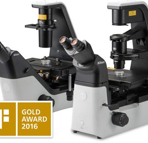
Inverted microscopes ECLIPSE Ts2 and ECLIPSE Ts2R recognized for streamlined body design
Nikon Instruments Inc. is pleased to announce that Inverted Microscopes ECLIPSE Ts2R and ECLIPSE Ts2 have been recognized with the iF Gold Award, a globally prestigious design recognition sponsored by iF International Forum Design GmbH since 1953. The ECLIPSE Ts2R and ECLIPSE Ts2 were awarded in the Product category (Industry/Skilled Trades) for their streamlined, space-saving design, ergonomics and enhanced functionality.
This year, 5,295 product and service entries from 53 countries were tested and evaluated by the iF Design Award judging panel, with 1,821 products receiving awards in the various categories. Of the winning products, 75 were given the iF Gold Award.
According to the iF jury “the ECLIPSE Ts2/Ts2R is a family of two microscopes with an unmistakable affiliation. A product that, due to its complexity, is very hard to design, has been implemented with amazing success. For designers it is a huge challenge to transfer this level of complexity into such a strong and clear design statement. Every detail is under control.”
Both the ECLIPSE Ts2R and ECLIPSE Ts2 systems feature Nikon’s latest design enhancements to improve observation, efficiency and accuracy.
- Improved body design: The compact, stable and durable body on both models conserves valuable bench space and easily fits next to an incubator or inside a tissue culture hood.
- Streamlined, ergonomic operation: The microscope can be conveniently switched between diascopic and epi-fluorescence observation using intuitively positioned controls and the lowered stage height of the Ts2R reduces fatigues during operation and frequent exchange of samples.
- LED-based diascopic and epi-fluorescence illumination: Eliminates alignment and frequent bulb replacements while providing bright and even illumination
- New Emboss Contrast technique: New cost-effective contrasting technique provides pseudo-three dimensional images of thick samples and is compatible with a variety of culture-ware including plastic dishes.
The ECLIPSE Ts2R and ECLIPSE Ts2 are now available for purchase.
ABOUT ECLIPSE Ts2:
Providing more accuracy and efficiency, the Inverted Routine Microscope ECLIPSE Ts2 is the successor of the ECLIPSE TS100. As an entry-level inverted microscope with improved functionality, it enables for more efficient microscopy while inheriting the highly reliable optical performance of the previous model.
Both the Ts2 and Ts2-FL models also employ LED illumination, eliminating the need for conventional adjustment and frequent replacement of light for both the diascopic and episcopic illuminators. Moreover, the zero warm-up time of LED illuminators allows for quicker cell observation.
ABOUT ECLIPSE Ts2R:
The ECLIPSE Ts2R is a compact inverted research microscope that is configurable with a wide variety of observation methods. The broad range of observation methods, smaller body and improved ease of use will directly benefit research laboratories and improve workflow.
In addition to conventional contrast methods, the ECLIPSE Ts2R features versatile observation methods, including Phase Contrast, DIC, Hoffman Modulation Contrast and Emboss Contrast. It is also compatible with a selection of high performance and quality optical accessories to function similarly to the inverted research microscope ECLIPSE Ti and produce clear, sharp images.
paramount March 3rd, 2016
Posted In: News Items
Olympus CX23 Microscope Offers Ruggedness, Dependability, Optical Excellence for Education, Training, Laboratory Use
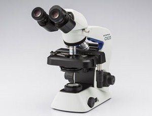
The CX23 educational microscope is easy to set up, operate and carry, making it perfect for use both by medical students and in laboratory operations that take place in the field and in other nontraditional settings. Built for rugged dependability over years of use, it weighs just 5.9kg (13 pounds), making it the lightest microscope in its class.
Like other instruments in the Olympus CX series, the new microscope features high-performance infinity-corrected optics for bright, clear, crisp images. The stable, color-corrected LED light source delivers reliable, natural-looking illumination, using far less power and lasting many times longer than traditional bulbs. A field number of 20 provides for added efficiency. The user can lock the frame to prevent any possibility of the objective and glass slide coming in contact, helping preventing accidental specimen damage.
The CX series is part of the full range Olympus scientific microscopes for education, clinical consultation, training and research. All are designed to deliver on the Olympus promise of optical excellence, performance and durability. From the flagship FLUOVIEW series of laser scanning confocal and multiphoton microscopes used in leading life science research facilities, through the powerhouse BX3/IX3 series that enable a wide range of live cell imaging applications, and on to the CX series for teaching in universities, field training sites and other educational institutions, Olympus provides microscopy solutions that exceed expectations for the widest variety of applications.
paramount April 2nd, 2015
Posted In: News Items